

What Is The Function Of A Mitochondria In A Animal Cell
Mitochondria
Mitochondria are rod-shaped organelles that can be considered the power generators of the jail cell, converting oxygen and nutrients into adenosine triphosphate (ATP). ATP is the chemic free energy "currency" of the cell that powers the cell'southward metabolic activities. This process is called aerobic respiration and is the reason animals exhale oxygen. Without mitochondria (singular, mitochondrion), higher animals would likely not exist because their cells would only be able to obtain energy from anaerobic respiration (in the absence of oxygen), a procedure much less efficient than aerobic respiration. In fact, mitochondria enable cells to produce 15 times more than ATP than they could otherwise, and complex animals, like humans, need large amounts of free energy in social club to survive.
The number of mitochondria present in a cell depends upon the metabolic requirements of that cell, and may range from a single large mitochondrion to thousands of the organelles. Mitochondria, which are found in most all eukaryotes, including plants, animals, fungi, and protists, are large enough to be observed with a light microscope and were starting time discovered in the 1800s. The proper name of the organelles was coined to reflect the way they looked to the first scientists to find them, stemming from the Greek words for "thread" and "granule." For many years subsequently their discovery, mitochondria were unremarkably believed to transmit hereditary data. It was not until the mid-1950s when a method for isolating the organelles intact was adult that the mod understanding of mitochondrial function was worked out.
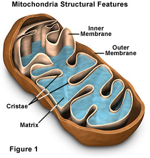
The elaborate construction of a mitochondrion is very of import to the functioning of the organelle (see Figure ane). 2 specialized membranes encircle each mitochondrion present in a prison cell, dividing the organelle into a narrow intermembrane space and a much larger internal matrix, each of which contains highly specialized proteins. The outer membrane of a mitochondrion contains many channels formed by the poly peptide porin and acts similar a sieve, filtering out molecules that are as well big. Similarly, the inner membrane, which is highly convoluted so that a large number of infoldings called cristae are formed, as well allows but sure molecules to pass through it and is much more selective than the outer membrane. To make certain that only those materials essential to the matrix are allowed into it, the inner membrane utilizes a grouping of transport proteins that will but send the correct molecules. Together, the diverse compartments of a mitochondrion are able to work in harmony to generate ATP in a complex multi-pace process.
Mitochondria are generally oblong organelles, which range in size between 1 and 10 micrometers in length, and occur in numbers that straight correlate with the cell's level of metabolic action. The organelles are quite flexible, however, and time-lapse studies of living cells have demonstrated that mitochondria change shape rapidly and motility about in the cell near constantly. Movements of the organelles appear to be linked in some manner to the microtubules present in the cell, and are probably transported along the network with motor proteins. Consequently, mitochondria may be organized into lengthy traveling chains, packed tightly into relatively stable groups, or appear in many other formations based upon the particular needs of the jail cell and the characteristics of its microtubular network.
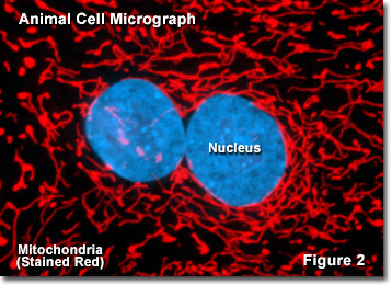
Presented in Figure 2 is a digital image of the mitochondrial network establish in the ovarian tissue from a mountain caprine animal relative, known as the Himalayan Tahr, as seen through a fluorescence optical microscope. The all-encompassing intertwined network is labeled with a synthetic dye named MitoTracker Scarlet (ruby-red fluorescence) that localizes in the respiring mitochondria of living cells in culture. The rare twin nuclei in this cell were counterstained with a blueish dye (cyan fluorescence) to denote their centralized location in relation to the mitochondrial network. Fluorescence microscopy is an important tool that scientists apply to examine the structure and function of internal cellular organelles.
The mitochondrion is unlike from most other organelles because it has its own circular DNA (like to the DNA of prokaryotes) and reproduces independently of the cell in which it is found; an apparent case of endosymbiosis. Scientists hypothesize that millions of years ago small, free-living prokaryotes were engulfed, but not consumed, by larger prokaryotes, mayhap because they were able to resist the digestive enzymes of the host organism. The two organisms adult a symbiotic human relationship over time, the larger organism providing the smaller with aplenty nutrients and the smaller organism providing ATP molecules to the larger one. Eventually, according to this view, the larger organism developed into the eukaryotic cell and the smaller organism into the mitochondrion.
Mitochondrial DNA is localized to the matrix, which also contains a host of enzymes, as well every bit ribosomes for protein synthesis. Many of the disquisitional metabolic steps of cellular respiration are catalyzed by enzymes that are able to diffuse through the mitochondrial matrix. The other proteins involved in respiration, including the enzyme that generates ATP, are embedded within the mitochondrial inner membrane. Infolding of the cristae dramatically increases the surface surface area available for hosting the enzymes responsible for cellular respiration.
Mitochondria are similar to institute chloroplasts in that both organelles are able to produce free energy and metabolites that are required by the host jail cell. Equally discussed above, mitochondria are the sites of respiration, and generate chemical energy in the form of ATP past metabolizing sugars, fats, and other chemic fuels with the assistance of molecular oxygen. Chloroplasts, in contrast, are institute merely in plants and algae, and are the main sites of photosynthesis. These organelles work in a different manner to convert energy from the sun into the biosynthesis of required organic nutrients using carbon dioxide and h2o. Similar mitochondria, chloroplasts likewise contain their own DNA and are able to grow and reproduce independently inside the cell.
In most animal species, mitochondria appear to be primarily inherited through the maternal lineage, though some contempo bear witness suggests that in rare instances mitochondria may also be inherited via a paternal route. Typically, a sperm carries mitochondria in its tail every bit an energy source for its long journey to the egg. When the sperm attaches to the egg during fertilization, the tail falls off. Consequently, the only mitochondria the new organism ordinarily gets are from the egg its mother provided. Therefore, different nuclear DNA, mitochondrial DNA doesn't get shuffled every generation, so it is presumed to change at a slower rate, which is useful for the report of man evolution. Mitochondrial Deoxyribonucleic acid is also used in forensic scientific discipline as a tool for identifying corpses or body parts, and has been implicated in a number of genetic diseases, such as Alzheimer'south disease and diabetes.
Dorsum TO ANIMAL CELL Construction
Back TO PLANT Cell STRUCTURE
Questions or comments? Transport us an email.
© 1995-2022 by Michael W. Davidson and The Florida State University. All Rights Reserved. No images, graphics, software, scripts, or applets may be reproduced or used in whatsoever manner without permission from the copyright holders. Employ of this website means y'all agree to all of the Legal Terms and Weather condition prepare along by the owners.
This website is maintained past our
Graphics & Web Programming Team
in collaboration with Optical Microscopy at the
National High Magnetic Field Laboratory.
Last modification: Friday, November 13, 2015 at 02:eighteen PM
Access Count Since October 1, 2000: 2214936
Microscopes provided by:
Source: https://micro.magnet.fsu.edu/cells/mitochondria/mitochondria.html
Posted by: murphyroyshe.blogspot.com
0 Response to "What Is The Function Of A Mitochondria In A Animal Cell"
Post a Comment